
Sovereign Diagnostic Tracking: Evaluating PET MRI CT Records and the NLR ctDNA Structural Biomarker Analysis

Diagnostics for Analysts: Evaluating Blood Test Biomarkers, CT, PET, and MRI Scans From a Patient Perspective to Facilitate Collaborative Discussions With the Primary Oncologist
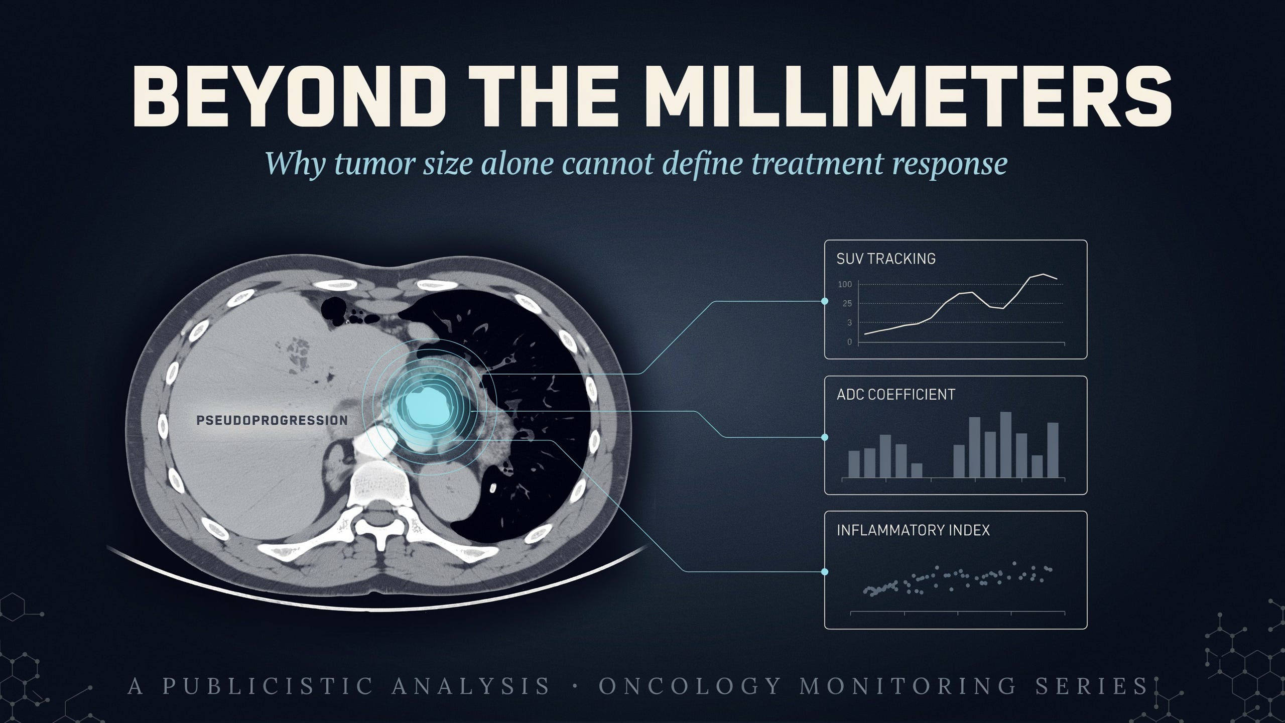
This publicistic article contains a comprehensive evaluation of key radiological parameters and laboratory indicators that directly outline the current status of oncological processes within the host organism. Independent tracking and the accurate interpretation of medical documentation remain exceptionally important because understanding the true biological baseline prevents chaotic decision timelines driven entirely by emotional strain. The circulating scientific literature demonstrates unequivocally that the physical macro dimensions of a tumor recorded in millimeters do not reflect the complete intracellular reality of the malignant matrix. During the active deployment of advanced metabolic protocols, the physical volume of a tumor can experience a temporary expansion due to intense immune cell infiltration and localized inflammatory edema, clinically defined within radiology as pseudoprogression. Therefore the systematic verification of digital parameters including Hounsfield units, ADC coefficients, and SUV tracking values remains a cornerstone requirement to evaluate the actual cellular viability of aggressive lineages. This objective publicistic analysis delivers structured research guidelines to sovereign families, facilitating highly technical and collaborative discussions alongside the primary oncology team.
Correlating the exact dynamics of tumor specific surface antigens concurrently with systemic inflammatory indicators establishes the bedrock foundation of modern diagnostic monitoring. Aggressive lineages circulating within advanced stage 4 presentations continuously stimulate localized inflammatory cascades within the tumor microenvironment, directly altering baseline hematological and biochemical serum values. Relying exclusively on standard reference boundaries printed on laboratory layouts remains insufficient because single values isolate individual markers, ignoring the complex biological interactions between host immunity and tumor mass structures. The compiled academic data confirms that monitoring advanced off label therapies requires a continuous cross matching strategy pairing tissue destruction tracking alongside unyielding density evaluations via PET and MRI imaging. Implementing a precise engineering perspective enables a reliable differentiation between genuine disease progression tracks and transient inflammatory waves triggered by the massive cellular disintegration of tumor fragments. Replacing the standard physiological dread surrounding complex medical terminology with an objective laboratory analysis allows for complete control over the therapeutic timeline.
Table of Contents
Section One: The Anatomy of Computed Tomography CT Scans: Evaluating Tissue Density and the Nephrotoxic Complications of Contrast Media
Section Two: The Functional Evaluation of PET Imaging: Interpreting SUV Metabolic Indices and Glucose Uptake Velocities
Section Three: The Specificity of Magnetic Resonance Imaging MRI: Evaluating Soft Tissue Integrity and Neurological Barriers
Section Four: Tumor Specific Antigens Versus Systemic Inflammation: Integrating the Neutrophil to Lymphocyte Ratio NLR Matrix
Section Five: Liquid Biopsy Frameworks: The Analytical Superiority of Circulating Tumor DNA ctDNA Tracking Arrays
Section Six: The Dynamics of Tumor Lysis Syndrome: Why Temporary Biomarker Spikes Signal Therapeutic Success
Section Seven: Tactical Consultation Preparation: Organizing Data Arrays to Facilitate Equal Peer Level Discussions
Section Eight: Academic Reference Repository and Clinical Biomarker Bibliography